| [1] |
FERNANDES K , CHICCO D , CARDOSO J S ,et al. Supervised deep learning embeddings for the prediction of cervical cancer diagnosis[J]. PeerJ Computer Science, 2018,4:e154.
|
| [2] |
SUN H F , YANG J H , FAN R B ,et al. Stepwise local stitching ultrasound image algorithms based on adaptive iterative threshold harris corner features[J]. Medicine, 2020,99(37): e22189.
|
| [3] |
HSU W Y , LU C C , HSU Y Y . Improving segmentation accuracy of CT kidney cancer images using adaptive active contour model[J]. Medicine, 2020,99(47): e23083.
|
| [4] |
何安良, 程兴保, 廖龙长 ,等. 耦合H-minima与数学形态学的分水岭遥感图像分割方法[J]. 东华理工大学学报(自然科学版), 2020,43(4): 396-400.
|
|
HE A L , CHENG X B , LIAO L C ,et al. An improved watershed method for remote sensing image segmentation coupling H-minima with mathematical morphology[J]. Journal of East China University of Technology (Natural Science), 2020,43(4): 396-400.
|
| [5] |
CHO M . Performance comparison of two ellipse fitting-based cell separation algorithms[J]. Journal of Information and Communication Convergence Engineering, 2015,13(3): 215-219.
|
| [6] |
KOYUNCU C F , AKHAN E , ERSAHIN T ,et al. Iterative H-minima-based marker-controlled watershed for cell nucleus segmentation[J]. Cytometry Part A, 2016,89(4): 338-349.
|
| [7] |
廖苗, 赵于前, 曾业战 ,等. 基于支持向量机和椭圆拟合的细胞图像自动分割[J]. 浙江大学学报(工学版), 2017,51(4): 722-728.
|
|
LIAO M , ZHAO Y Q , ZENG Y Z ,et al. Automatic segmentation for cell images based on support vector machine and ellipse fitting[J]. Journal of Zhejiang University (Engineering Science), 2017,51(4): 722-728.
|
| [8] |
杨秀杰, 李法平 . 基于曲率和活动轮廓模型的重叠细胞分割算法[J]. 西南师范大学学报(自然科学版), 2018,43(4): 41-47.
|
|
YANG X J , LI F P . Overlapping cells segmentation algorithm based on curvature and active contour model[J]. Journal of Southwest China Normal University (Natural Science Edition), 2018,43(4): 41-47.
|
| [9] |
何国生, 施露露, 邹爽爽 ,等. 基于自适应阈值的间充质干细胞分割方法研究[J]. 电子测量与仪器学报, 2019,33(6): 18-23.
|
|
HE G S , SHI L L , ZOU S S ,et al. Research on mesenchymal stem cells segmentation based on adaptive threshold[J]. Journal of Electronic Measurement and Instrumentation, 2019,33(6): 18-23.
|
| [10] |
FABIJA?SKA A . Segmentation of corneal endothelium images using a U-Net-based convolutional neural network[J]. Artificial Intelligence in Medicine, 2018,88: 1-13.
|
| [11] |
NASR-ESFAHANI E , RAFIEI S , JAFARI M H ,et al. Dense pooling layers in fully convolutional network for skin lesion segmentation[J]. Computerized Medical Imaging and Graphics, 2019,78:101658.
|
| [12] |
SAQIB Q , HAI J , RAN Z ,et al. A variant form of 3D-UNet for infant brain segmentation[J]. Future Generation Computer Systems, 2020,108(7): 613-623.
|
| [13] |
SHIMAA E-B , AHMAD A-K , MAHA S . A two-stage framework for automated malignant pulmonary nodule detection in CT scans[J]. Diagnostics, 2020,10(3): e131.
|
| [14] |
LIU Z . Construction and verification of color fundus image retinal vessels segmentation algorithm under BP neural network[J]. The Journal of Supercomputing, 2021,77(7): 7171-7183.
|
| [15] |
崔文成, 张鹏霞, 邵虹 . 基于深度可分离卷积网络的皮肤镜图像病灶分割方法[J]. 智能科学与技术学报, 2020,2(4): 385-393.
|
|
CUI W C , ZHANG P X , SHAO H . Dermoscopic image lesion segmentation method based on deep separable convolutional network[J]. Chinese Journal of Intelligent Science and Technology, 2020,2(4): 385-393.
|
| [16] |
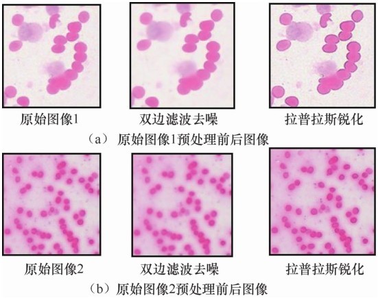
王涛, 陈凡胜, 苏晓锋 . 基于各向异性双边滤波红外背景抑制方法研究[J]. 湖南大学学报(自然科学版), 2018,45(2): 119-126.
|
|
WANG T , CHEN F S , SU X F . Research of infrared background suppression method based on anisotropic bilateral filtering[J]. Journal of Hunan University (Natural Sciences), 2018,45(2): 119-126.
|
| [17] |
王浩, 张叶, 沈宏海 ,等. 图像增强算法综述[J]. 中国光学, 2017,10(4): 438-448.
|
|
WANG H , ZHANG Y , SHEN H H ,et al. Review of image enhancement algorithms[J]. Chinese Optics, 2017,10(4): 438-448.
|
| [18] |
OKTAY O , SCHLEMPER J , FOLGOC L L ,et al. Attention U-Net:learning where to look for the pancreas[J]. arXiv preprint, 2018,arXiv:1804.03999.
|
| [19] |
WANG H , ZHANG H , RAY N . Clump splitting via bottleneck detection and shape classification[J]. Pattern Recognition, 2012,45(7): 2780-2787.
|