Cognition and depression:current status and future directions
1
2010
... 抑郁症是一种常见的精神疾病,其表现为心境低落、思维迟缓、意志活动减退、认知功能损害等[1],且具有高发病率[2]、高复发率[3]、高自杀率[4]等特点,严重危害患者的身心健康.抑郁症已成为世界四大疾病之一,目前已有3.5亿患者,并将在2020年成为世界第二大疾病[5].其发病因素众多,患者受生物、心理以及社会环境等多个方面的影响,而关于抑郁症的发病机制仍在研究中. ...
Lifetime prevalence and age-of-onset distributions of DSM-IV disorders in the national comorbidity survey replication
1
2005
... 抑郁症是一种常见的精神疾病,其表现为心境低落、思维迟缓、意志活动减退、认知功能损害等[1],且具有高发病率[2]、高复发率[3]、高自杀率[4]等特点,严重危害患者的身心健康.抑郁症已成为世界四大疾病之一,目前已有3.5亿患者,并将在2020年成为世界第二大疾病[5].其发病因素众多,患者受生物、心理以及社会环境等多个方面的影响,而关于抑郁症的发病机制仍在研究中. ...
The prospective long-term course of adult depression in general practice and the community:a systematic literature review
1
2014
... 抑郁症是一种常见的精神疾病,其表现为心境低落、思维迟缓、意志活动减退、认知功能损害等[1],且具有高发病率[2]、高复发率[3]、高自杀率[4]等特点,严重危害患者的身心健康.抑郁症已成为世界四大疾病之一,目前已有3.5亿患者,并将在2020年成为世界第二大疾病[5].其发病因素众多,患者受生物、心理以及社会环境等多个方面的影响,而关于抑郁症的发病机制仍在研究中. ...
Can better recognition and treatment of depression reduce suicide rates? A brief review
1
2001
... 抑郁症是一种常见的精神疾病,其表现为心境低落、思维迟缓、意志活动减退、认知功能损害等[1],且具有高发病率[2]、高复发率[3]、高自杀率[4]等特点,严重危害患者的身心健康.抑郁症已成为世界四大疾病之一,目前已有3.5亿患者,并将在2020年成为世界第二大疾病[5].其发病因素众多,患者受生物、心理以及社会环境等多个方面的影响,而关于抑郁症的发病机制仍在研究中. ...
Evidence-based health policy-lessons from the global burden of disease study
1
1996
... 抑郁症是一种常见的精神疾病,其表现为心境低落、思维迟缓、意志活动减退、认知功能损害等[1],且具有高发病率[2]、高复发率[3]、高自杀率[4]等特点,严重危害患者的身心健康.抑郁症已成为世界四大疾病之一,目前已有3.5亿患者,并将在2020年成为世界第二大疾病[5].其发病因素众多,患者受生物、心理以及社会环境等多个方面的影响,而关于抑郁症的发病机制仍在研究中. ...
The rise and fall of MRI studies in major depressive disorder
1
2019
... 对抑郁症的诊断目前主要依赖于临床症状,综合严重程度以及症状数量将患者划分为轻度抑郁症、中度抑郁症和重度抑郁症.近年来,随着磁共振影像技术的发展,出现了大量关于抑郁症患者大脑结构以及功能异常的研究,对抑郁症的病理变化及致病机制有了一定的了解[6,7],通过对脑影像进行分析,可以辅助医生做出更准确的诊断、早期预测以及干预;通过分析不同服药阶段的表现来评估药物疗效.磁共振成像主要分为两大类:结构性磁共振成像(structural magnetic resonance imaging, sMRI)和功能性磁共振成像(functional magnetic resonance imaging,fMRI),这些成像方法对人体均无创.抑郁症患者大脑结构形态学的显著变化出现在颞叶、前扣带皮层、眶额叶皮层、海马体、杏仁核、丘脑等区域[8,9],皮层的表面积和厚度都发生了显著变化,且皮下各结构出现了不同程度的萎缩和膨胀,这些结构都与情绪调节以及记忆等功能相关.而在结构网络和功能网络方面,连接的组织效率、连通性都出现了降低,小世界属性丧失,拓扑属性遭到破坏.抑郁症影像学的研究可应用于临床的辅助诊断、药物治疗效果评估、高危人群预测等.本文简述了不同影像的分析方法,并详细探讨了与抑郁症相关的3个重要的功能子网络,结合当前研究现状对未来的研究提出了建议. ...
Major depressive disorder and magnetic resonance imaging:a mini-review of recent progress
1
2018
... 对抑郁症的诊断目前主要依赖于临床症状,综合严重程度以及症状数量将患者划分为轻度抑郁症、中度抑郁症和重度抑郁症.近年来,随着磁共振影像技术的发展,出现了大量关于抑郁症患者大脑结构以及功能异常的研究,对抑郁症的病理变化及致病机制有了一定的了解[6,7],通过对脑影像进行分析,可以辅助医生做出更准确的诊断、早期预测以及干预;通过分析不同服药阶段的表现来评估药物疗效.磁共振成像主要分为两大类:结构性磁共振成像(structural magnetic resonance imaging, sMRI)和功能性磁共振成像(functional magnetic resonance imaging,fMRI),这些成像方法对人体均无创.抑郁症患者大脑结构形态学的显著变化出现在颞叶、前扣带皮层、眶额叶皮层、海马体、杏仁核、丘脑等区域[8,9],皮层的表面积和厚度都发生了显著变化,且皮下各结构出现了不同程度的萎缩和膨胀,这些结构都与情绪调节以及记忆等功能相关.而在结构网络和功能网络方面,连接的组织效率、连通性都出现了降低,小世界属性丧失,拓扑属性遭到破坏.抑郁症影像学的研究可应用于临床的辅助诊断、药物治疗效果评估、高危人群预测等.本文简述了不同影像的分析方法,并详细探讨了与抑郁症相关的3个重要的功能子网络,结合当前研究现状对未来的研究提出了建议. ...
Subcortical brain alterations in major depressive disorder:findings from the enigma major depressive disorder working group
2
2016
... 对抑郁症的诊断目前主要依赖于临床症状,综合严重程度以及症状数量将患者划分为轻度抑郁症、中度抑郁症和重度抑郁症.近年来,随着磁共振影像技术的发展,出现了大量关于抑郁症患者大脑结构以及功能异常的研究,对抑郁症的病理变化及致病机制有了一定的了解[6,7],通过对脑影像进行分析,可以辅助医生做出更准确的诊断、早期预测以及干预;通过分析不同服药阶段的表现来评估药物疗效.磁共振成像主要分为两大类:结构性磁共振成像(structural magnetic resonance imaging, sMRI)和功能性磁共振成像(functional magnetic resonance imaging,fMRI),这些成像方法对人体均无创.抑郁症患者大脑结构形态学的显著变化出现在颞叶、前扣带皮层、眶额叶皮层、海马体、杏仁核、丘脑等区域[8,9],皮层的表面积和厚度都发生了显著变化,且皮下各结构出现了不同程度的萎缩和膨胀,这些结构都与情绪调节以及记忆等功能相关.而在结构网络和功能网络方面,连接的组织效率、连通性都出现了降低,小世界属性丧失,拓扑属性遭到破坏.抑郁症影像学的研究可应用于临床的辅助诊断、药物治疗效果评估、高危人群预测等.本文简述了不同影像的分析方法,并详细探讨了与抑郁症相关的3个重要的功能子网络,结合当前研究现状对未来的研究提出了建议. ...
... 不同年龄的抑郁症患者有不同的表现,成年重度抑郁症患者形态变化主要为皮层厚度变薄,青少年重度抑郁症患者表现为大脑皮层表面积减少[9],晚发型抑郁症患者的皮下结构变化程度明显比早发型抑郁症患者的皮下结构变化程度高.而对于不同类型的抑郁症来说,复发型抑郁症患者的大脑变化要显著大于首发型抑郁症患者的大脑变化[8].同时,治疗的不同阶段也对应不同的大脑异常变化. ...
Cortical abnormalities in adults and adolescents with major depression based on brain scans from 20 cohorts worldwide in the enigma major depressive disorder working group
2
2017
... 对抑郁症的诊断目前主要依赖于临床症状,综合严重程度以及症状数量将患者划分为轻度抑郁症、中度抑郁症和重度抑郁症.近年来,随着磁共振影像技术的发展,出现了大量关于抑郁症患者大脑结构以及功能异常的研究,对抑郁症的病理变化及致病机制有了一定的了解[6,7],通过对脑影像进行分析,可以辅助医生做出更准确的诊断、早期预测以及干预;通过分析不同服药阶段的表现来评估药物疗效.磁共振成像主要分为两大类:结构性磁共振成像(structural magnetic resonance imaging, sMRI)和功能性磁共振成像(functional magnetic resonance imaging,fMRI),这些成像方法对人体均无创.抑郁症患者大脑结构形态学的显著变化出现在颞叶、前扣带皮层、眶额叶皮层、海马体、杏仁核、丘脑等区域[8,9],皮层的表面积和厚度都发生了显著变化,且皮下各结构出现了不同程度的萎缩和膨胀,这些结构都与情绪调节以及记忆等功能相关.而在结构网络和功能网络方面,连接的组织效率、连通性都出现了降低,小世界属性丧失,拓扑属性遭到破坏.抑郁症影像学的研究可应用于临床的辅助诊断、药物治疗效果评估、高危人群预测等.本文简述了不同影像的分析方法,并详细探讨了与抑郁症相关的3个重要的功能子网络,结合当前研究现状对未来的研究提出了建议. ...
... 不同年龄的抑郁症患者有不同的表现,成年重度抑郁症患者形态变化主要为皮层厚度变薄,青少年重度抑郁症患者表现为大脑皮层表面积减少[9],晚发型抑郁症患者的皮下结构变化程度明显比早发型抑郁症患者的皮下结构变化程度高.而对于不同类型的抑郁症来说,复发型抑郁症患者的大脑变化要显著大于首发型抑郁症患者的大脑变化[8].同时,治疗的不同阶段也对应不同的大脑异常变化. ...
Validation of freesurfer-estimated brain cortical thickness:comparison with histologic measurements
1
2014
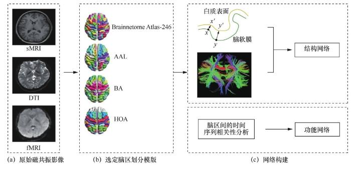
... 形态学分析主要基于皮层厚度、体积、表面积、密度等属性,从解剖学的角度直观反映大脑的变化,其中皮质厚度较难直接测量,因此将其定义为白质表面和软膜表面的最短距离,该方法的有效性已得到验证[10].除形态学分析之外,大部分研究集中在脑网络分析,使用结构连接或者功能连接构建网络,对抑郁症患者的大脑变化进一步分析,其中每个感兴趣区域(region of interest,ROI)代表一个节点,每个感兴趣区域有其特定的功能,各感兴趣区域之间协同工作. ...
Vergleichende lokalisationslehre der grobhirnrinde
1
1909
... 结构网络基于sMRI进行构建,反映了大脑结构连通性的异常,通常使用脑影像分析工具FreeSurfer 或统计参数图(statistical parametric mapping,SPM)等来计算大脑皮层厚度或灰质体积,根据选定脑区模板进行感兴趣区域的划分.基于每个感兴趣区域的皮层厚度或灰质体积构建结构网络,常用模板有Brodmann(BA)[11]、自动解剖标签(automated anatomical labeling,AAL)[12]、Harvard-Oxford Atlas(HOA)[13]、Brainnetome Atlas-246 [14]等.使用皮尔逊相关性分析来构建各脑区之间的连接,若选择的模板为n个脑区,则形成n×n 的连接矩阵,矩阵的每一行或每一列均表示一个脑区.基于脑网络的拓扑属性分析包括全局或局部属性、模块化、节点属性等,如节点强度表示节点的连接数,聚类系数表示某一节点i的相邻节点之间也互为相邻节点的可能性.其中,中心度用于度量网络中节点的作用与地位,最短路径长度与全局效率衡量了网络的全局传输能力,各项指标既包含大脑整体的完整性,也涉及各功能脑区的变化. ...
Automated anatomical labeling of activations in SPM using a macroscopic anatomical parcellation of the MNI MRI single-subject brain
1
2002
... 结构网络基于sMRI进行构建,反映了大脑结构连通性的异常,通常使用脑影像分析工具FreeSurfer 或统计参数图(statistical parametric mapping,SPM)等来计算大脑皮层厚度或灰质体积,根据选定脑区模板进行感兴趣区域的划分.基于每个感兴趣区域的皮层厚度或灰质体积构建结构网络,常用模板有Brodmann(BA)[11]、自动解剖标签(automated anatomical labeling,AAL)[12]、Harvard-Oxford Atlas(HOA)[13]、Brainnetome Atlas-246 [14]等.使用皮尔逊相关性分析来构建各脑区之间的连接,若选择的模板为n个脑区,则形成n×n 的连接矩阵,矩阵的每一行或每一列均表示一个脑区.基于脑网络的拓扑属性分析包括全局或局部属性、模块化、节点属性等,如节点强度表示节点的连接数,聚类系数表示某一节点i的相邻节点之间也互为相邻节点的可能性.其中,中心度用于度量网络中节点的作用与地位,最短路径长度与全局效率衡量了网络的全局传输能力,各项指标既包含大脑整体的完整性,也涉及各功能脑区的变化. ...
Advances in functional and structural MR image analysis and implementation as FSL
1
2004
... 结构网络基于sMRI进行构建,反映了大脑结构连通性的异常,通常使用脑影像分析工具FreeSurfer 或统计参数图(statistical parametric mapping,SPM)等来计算大脑皮层厚度或灰质体积,根据选定脑区模板进行感兴趣区域的划分.基于每个感兴趣区域的皮层厚度或灰质体积构建结构网络,常用模板有Brodmann(BA)[11]、自动解剖标签(automated anatomical labeling,AAL)[12]、Harvard-Oxford Atlas(HOA)[13]、Brainnetome Atlas-246 [14]等.使用皮尔逊相关性分析来构建各脑区之间的连接,若选择的模板为n个脑区,则形成n×n 的连接矩阵,矩阵的每一行或每一列均表示一个脑区.基于脑网络的拓扑属性分析包括全局或局部属性、模块化、节点属性等,如节点强度表示节点的连接数,聚类系数表示某一节点i的相邻节点之间也互为相邻节点的可能性.其中,中心度用于度量网络中节点的作用与地位,最短路径长度与全局效率衡量了网络的全局传输能力,各项指标既包含大脑整体的完整性,也涉及各功能脑区的变化. ...
The human brainnetome atlas:a new brain atlas based on connectional architecture
1
2016
... 结构网络基于sMRI进行构建,反映了大脑结构连通性的异常,通常使用脑影像分析工具FreeSurfer 或统计参数图(statistical parametric mapping,SPM)等来计算大脑皮层厚度或灰质体积,根据选定脑区模板进行感兴趣区域的划分.基于每个感兴趣区域的皮层厚度或灰质体积构建结构网络,常用模板有Brodmann(BA)[11]、自动解剖标签(automated anatomical labeling,AAL)[12]、Harvard-Oxford Atlas(HOA)[13]、Brainnetome Atlas-246 [14]等.使用皮尔逊相关性分析来构建各脑区之间的连接,若选择的模板为n个脑区,则形成n×n 的连接矩阵,矩阵的每一行或每一列均表示一个脑区.基于脑网络的拓扑属性分析包括全局或局部属性、模块化、节点属性等,如节点强度表示节点的连接数,聚类系数表示某一节点i的相邻节点之间也互为相邻节点的可能性.其中,中心度用于度量网络中节点的作用与地位,最短路径长度与全局效率衡量了网络的全局传输能力,各项指标既包含大脑整体的完整性,也涉及各功能脑区的变化. ...
Multimodal imaging reveals a complex pattern of dysfunction in corticolimbic pathways in major depressive disorder
1
2019
... 除此之外,弥散张量成像(diffusion tensor imaging,DTI)数据也常被用于构建结构网络,DTI网络通过对全脑纤维束进行追踪,从白质纤维角度评估大脑结构的变化,常用的度量包括平均弥散率(mean diffusivity,MD)和各向异性分数(fractional anisotropy,FA)等指标[15]. ...
Aberrant topological organization of the functional brain network associated with prior overt hepatic encephalopathy in cirrhotic patients
1
2019
... 除了局部一致性(regional homogeneity,ReHo)与低频振幅(amplitude of low frequency fluctuation, ALFF)2个属性,目前对静息态fMRI数据的分析常常基于先验模板或独立成分分析(independent component analysis,ICA)来提取感兴趣区域的血氧合水平依赖(blood oxygenation level dependent, Bold)信号,相比于先验模板提供已经制定好的脑区划分,ICA是一种数据驱动的信号处理方法,不同的研究可能提取出不同数量的独立成分,每个独立成分代表一个脑区.计算每个脑区的平均时间序列,对其进行皮尔逊相关性分析,通过 Fisher’s-z变换将数据转换为正态分布的z值,将计算的相关系数作为网络中边的权值,构建功能连接矩阵,并计算拓扑属性,功能网络的构建过程可借助已有的开源工具来实现,如 GRETNA 等[16,17].在网络属性的分析方面,功能网络与结构网络相似. ...
阿尔兹海默症脑网络演化建模
1
2019
... 除了局部一致性(regional homogeneity,ReHo)与低频振幅(amplitude of low frequency fluctuation, ALFF)2个属性,目前对静息态fMRI数据的分析常常基于先验模板或独立成分分析(independent component analysis,ICA)来提取感兴趣区域的血氧合水平依赖(blood oxygenation level dependent, Bold)信号,相比于先验模板提供已经制定好的脑区划分,ICA是一种数据驱动的信号处理方法,不同的研究可能提取出不同数量的独立成分,每个独立成分代表一个脑区.计算每个脑区的平均时间序列,对其进行皮尔逊相关性分析,通过 Fisher’s-z变换将数据转换为正态分布的z值,将计算的相关系数作为网络中边的权值,构建功能连接矩阵,并计算拓扑属性,功能网络的构建过程可借助已有的开源工具来实现,如 GRETNA 等[16,17].在网络属性的分析方面,功能网络与结构网络相似. ...
Instability of default mode network connectivity in major depression:a two-sample confirmation study
1
2017
... 大量关于抑郁症的研究表明,患者的大脑异常主要出现在三大功能子网络,包括默认模式网络(default mode network,DMN)[18,19]、认知控制网络(cognitive control network,CCN)[20,21]以及突显网络(salience network,SN)[20,21,22],且这三大功能子网络在调节情绪和认知功能上起到了极其重要的作用.在通过脑网络进行分析时,若研究目的是讨论某功能子网络的变化,那么在处理数据前只需挑选该功能子网络涉及的脑区即可,然后再构建网络. ...
Rumination and default mode network subsystems connectivity in first-episode,drug-naive young patients with major depressive disorder
2
2017
... 大量关于抑郁症的研究表明,患者的大脑异常主要出现在三大功能子网络,包括默认模式网络(default mode network,DMN)[18,19]、认知控制网络(cognitive control network,CCN)[20,21]以及突显网络(salience network,SN)[20,21,22],且这三大功能子网络在调节情绪和认知功能上起到了极其重要的作用.在通过脑网络进行分析时,若研究目的是讨论某功能子网络的变化,那么在处理数据前只需挑选该功能子网络涉及的脑区即可,然后再构建网络. ...
... 默认模式网络在大脑不进行任务时极其活跃,而抑郁症患者的DMN相比正常人更为活跃[26,27],前DMN功能连接增加,后DMN部分功能连接减少[28],DMN 整体连通性增加.有研究推测 DMN的连接异常与抑郁症的反刍思维存在关联[19]. ...
Altered functional interaction hub between affective network and cognitive control network in patients with major depressive disorder
2
2016
... 大量关于抑郁症的研究表明,患者的大脑异常主要出现在三大功能子网络,包括默认模式网络(default mode network,DMN)[18,19]、认知控制网络(cognitive control network,CCN)[20,21]以及突显网络(salience network,SN)[20,21,22],且这三大功能子网络在调节情绪和认知功能上起到了极其重要的作用.在通过脑网络进行分析时,若研究目的是讨论某功能子网络的变化,那么在处理数据前只需挑选该功能子网络涉及的脑区即可,然后再构建网络. ...
... [20,21,22],且这三大功能子网络在调节情绪和认知功能上起到了极其重要的作用.在通过脑网络进行分析时,若研究目的是讨论某功能子网络的变化,那么在处理数据前只需挑选该功能子网络涉及的脑区即可,然后再构建网络. ...
Comorbid anxiety increases cognitive control activation in major depressive disorder
2
2016
... 大量关于抑郁症的研究表明,患者的大脑异常主要出现在三大功能子网络,包括默认模式网络(default mode network,DMN)[18,19]、认知控制网络(cognitive control network,CCN)[20,21]以及突显网络(salience network,SN)[20,21,22],且这三大功能子网络在调节情绪和认知功能上起到了极其重要的作用.在通过脑网络进行分析时,若研究目的是讨论某功能子网络的变化,那么在处理数据前只需挑选该功能子网络涉及的脑区即可,然后再构建网络. ...
... ,21,22],且这三大功能子网络在调节情绪和认知功能上起到了极其重要的作用.在通过脑网络进行分析时,若研究目的是讨论某功能子网络的变化,那么在处理数据前只需挑选该功能子网络涉及的脑区即可,然后再构建网络. ...
Alteration of spontaneous neuronal activity within the salience network in partially remitted depression
1
2015
... 大量关于抑郁症的研究表明,患者的大脑异常主要出现在三大功能子网络,包括默认模式网络(default mode network,DMN)[18,19]、认知控制网络(cognitive control network,CCN)[20,21]以及突显网络(salience network,SN)[20,21,22],且这三大功能子网络在调节情绪和认知功能上起到了极其重要的作用.在通过脑网络进行分析时,若研究目的是讨论某功能子网络的变化,那么在处理数据前只需挑选该功能子网络涉及的脑区即可,然后再构建网络. ...
A treatment-resistant default mode subnetwork in major depression
1
2013
... DMN 可分为前 DMN(anterior DMN)和后DMN(posterior DMN)[23].前DMN主要以内侧前额叶皮层为中心,且与杏仁核相连,因此与自我参照处理(self-referential processing)和情绪调节有关,抑郁症患者的 DMN 变化主要发生在前 DMN[24];后DMN与海马体关联,主要与记忆和意识有关[25]. ...
The default network and self-generated thought:component processes,dynamic control,and clinical relevance
1
2014
... DMN 可分为前 DMN(anterior DMN)和后DMN(posterior DMN)[23].前DMN主要以内侧前额叶皮层为中心,且与杏仁核相连,因此与自我参照处理(self-referential processing)和情绪调节有关,抑郁症患者的 DMN 变化主要发生在前 DMN[24];后DMN与海马体关联,主要与记忆和意识有关[25]. ...
Resting-state functional connectivity in major depressive disorder:a review
2
2015
... DMN 可分为前 DMN(anterior DMN)和后DMN(posterior DMN)[23].前DMN主要以内侧前额叶皮层为中心,且与杏仁核相连,因此与自我参照处理(self-referential processing)和情绪调节有关,抑郁症患者的 DMN 变化主要发生在前 DMN[24];后DMN与海马体关联,主要与记忆和意识有关[25]. ...
... 突显网络对DMN和CCN的异常切换可能会导致抑郁症的产生,抑郁症患者突显网络与前 DMN之间的连通性增加,与 CCN 的连接减少,同时,其右前脑岛的内部连接显著减少[38].突显网络连接的异常程度与抑郁严重程度相关[25],因此推测右前脑岛的连接异常导致了DMN和CCN的切换异常. ...
Resting-state functional MRI in depression unmasks increased connectivity between networks via the dorsal nexus
1
2010
... 默认模式网络在大脑不进行任务时极其活跃,而抑郁症患者的DMN相比正常人更为活跃[26,27],前DMN功能连接增加,后DMN部分功能连接减少[28],DMN 整体连通性增加.有研究推测 DMN的连接异常与抑郁症的反刍思维存在关联[19]. ...
Self-referential processing,rumination,and cortical midline structures in major depression
1
2013
... 默认模式网络在大脑不进行任务时极其活跃,而抑郁症患者的DMN相比正常人更为活跃[26,27],前DMN功能连接增加,后DMN部分功能连接减少[28],DMN 整体连通性增加.有研究推测 DMN的连接异常与抑郁症的反刍思维存在关联[19]. ...
Resting state brain network function in major depression-depression symptomatology,antidepressant treatment effects,future research
1
2017
... 默认模式网络在大脑不进行任务时极其活跃,而抑郁症患者的DMN相比正常人更为活跃[26,27],前DMN功能连接增加,后DMN部分功能连接减少[28],DMN 整体连通性增加.有研究推测 DMN的连接异常与抑郁症的反刍思维存在关联[19]. ...
Cognitive control network anatomy correlates with neurocognitive behavior:a longitudinal study
1
2017
... 与默认模式网络在注意力和认知任务中休息相反,认知控制网络在认知任务中极其活跃,包括注意力、计划和工作记忆等[29].相较于健康个体,抑郁症患者认知控制网络的白质微结构异常,FA显著降低[30],功能连接与连通性显著降低[31],这些特征在与关注外部环境相关的区域[32]表现的尤为明显,已有研究发现认知控制网络的静态功能连接可预测药物治疗的预后[33]. ...
Serotonin transporter polymorphisms,microstructural white matter abnormalities and remission of geriatric depression
1
2009
... 与默认模式网络在注意力和认知任务中休息相反,认知控制网络在认知任务中极其活跃,包括注意力、计划和工作记忆等[29].相较于健康个体,抑郁症患者认知控制网络的白质微结构异常,FA显著降低[30],功能连接与连通性显著降低[31],这些特征在与关注外部环境相关的区域[32]表现的尤为明显,已有研究发现认知控制网络的静态功能连接可预测药物治疗的预后[33]. ...
Altered functional connectivity of the dorsolateral prefrontal cortex in first-episode patients with major depressive disorder
1
2012
... 与默认模式网络在注意力和认知任务中休息相反,认知控制网络在认知任务中极其活跃,包括注意力、计划和工作记忆等[29].相较于健康个体,抑郁症患者认知控制网络的白质微结构异常,FA显著降低[30],功能连接与连通性显著降低[31],这些特征在与关注外部环境相关的区域[32]表现的尤为明显,已有研究发现认知控制网络的静态功能连接可预测药物治疗的预后[33]. ...
Large-scale network dysfunction in major depressive disorder:a meta-analysis of resting-state functional connectivity
1
2015
... 与默认模式网络在注意力和认知任务中休息相反,认知控制网络在认知任务中极其活跃,包括注意力、计划和工作记忆等[29].相较于健康个体,抑郁症患者认知控制网络的白质微结构异常,FA显著降低[30],功能连接与连通性显著降低[31],这些特征在与关注外部环境相关的区域[32]表现的尤为明显,已有研究发现认知控制网络的静态功能连接可预测药物治疗的预后[33]. ...
Functional connectivity in the cognitive control network and the default mode network in late-life depression
1
2012
... 与默认模式网络在注意力和认知任务中休息相反,认知控制网络在认知任务中极其活跃,包括注意力、计划和工作记忆等[29].相较于健康个体,抑郁症患者认知控制网络的白质微结构异常,FA显著降低[30],功能连接与连通性显著降低[31],这些特征在与关注外部环境相关的区域[32]表现的尤为明显,已有研究发现认知控制网络的静态功能连接可预测药物治疗的预后[33]. ...
Dorsal anterior cingulate cortex shows fMRI response to internal and external error signals
1
2004
... 突显网络由双侧的前脑岛(anterior insula,AI)、背侧前扣带回(dorsal anterior cingulate cortex, dACC)、杏仁核以及颞极构成,其中dACC是认知控制中的关键结构[34,35]. ...
Cognitive control and the salience network:an investigation of error processing and effective connectivity
1
2013
... 突显网络由双侧的前脑岛(anterior insula,AI)、背侧前扣带回(dorsal anterior cingulate cortex, dACC)、杏仁核以及颞极构成,其中dACC是认知控制中的关键结构[34,35]. ...
The salience network is responsible for switching between the default mode network and the central executive network:replication from DCM
1
2014
... 突显网络主要协助大脑对收到的刺激做出反应,它的异常与抑郁症、精神分裂症等精神疾病显著相关,研究者发现了情绪、认知和平衡与突显网络的相关性.突显网络在DMN和CCN的切换中起到了重要作用[36,37],有研究发现突显网络中有一类被称为VEN(von economo neuron)的特殊神经元,该神经元可促进网络交换的过程,并且只在突显网络中被发现. ...
A critical role for the right fronto-insular cortex in switching between central-executive and default-mode networks
1
2008
... 突显网络主要协助大脑对收到的刺激做出反应,它的异常与抑郁症、精神分裂症等精神疾病显著相关,研究者发现了情绪、认知和平衡与突显网络的相关性.突显网络在DMN和CCN的切换中起到了重要作用[36,37],有研究发现突显网络中有一类被称为VEN(von economo neuron)的特殊神经元,该神经元可促进网络交换的过程,并且只在突显网络中被发现. ...
Insular dysfunction within the salience network is associated with severity of symptoms and aberrant inter-network connectivity in major depressive disorder
1
2014
... 突显网络对DMN和CCN的异常切换可能会导致抑郁症的产生,抑郁症患者突显网络与前 DMN之间的连通性增加,与 CCN 的连接减少,同时,其右前脑岛的内部连接显著减少[38].突显网络连接的异常程度与抑郁严重程度相关[25],因此推测右前脑岛的连接异常导致了DMN和CCN的切换异常. ...
Brain-derived neurotrophic factor promoter methylation and cortical thickness in recurrent major depressive disorder
2016
Paralimbic cortical thickness in first-episode depression:evidence for trait-related differences in mood regulation
2013
Regional increases of cortical thickness in untreated,first-episode major depressive disorder
2014
Widespread reductions in gray matter volume in depression
2013
Cerebral and cerebellar gray matter reduction in first-episode patients with major depressive disorder:a voxel-based morphometry study
1
2011
... 形态学的分析可从解剖学角度描述抑郁症患者的大脑变化,其中皮层厚度、表面积、灰质体积、密度为常讨论的特征.抑郁症患者的部分脑区皮层厚度显著变薄或变厚,其中异常较为显著地集中在双侧和内侧前额叶皮层、后扣带皮层、左侧颞中回、内侧眶额回等脑区[43,44],这些研究都体现了小脑在情绪调节以及记忆方面起到的作用. ...
Relationship between cerebellar structure and emotional memory in depression
1
2017
... 形态学的分析可从解剖学角度描述抑郁症患者的大脑变化,其中皮层厚度、表面积、灰质体积、密度为常讨论的特征.抑郁症患者的部分脑区皮层厚度显著变薄或变厚,其中异常较为显著地集中在双侧和内侧前额叶皮层、后扣带皮层、左侧颞中回、内侧眶额回等脑区[43,44],这些研究都体现了小脑在情绪调节以及记忆方面起到的作用. ...
Conscious and unconscious emotional learning in the human amygdala
1
1998
... 除了大脑皮层之外,抑郁症患者大脑形态学变化还出现在皮层下结构,发生显著变化的结构包括海马体、杏仁核、丘脑、壳核等,其中体积变化最大的是海马体.海马体和杏仁核在情绪调节[45]、表达[46]、工作记忆[47]等方面起到重要作用.不仅抑郁症患者的海马体发生了显著变化,精神分裂症(schizophrenia)[48]、阿尔兹海默症(Alzheimer’s disease,AD)[49]等精神疾病患者的海马体都发生了显著的变化.笔者研究分析了海马体和杏仁核不同子区的形态学变化,结果显示,双侧海马体的下托和安蒙角(cornu ammonis,CA)1子区,左侧杏仁核的外侧和基底外侧,右侧杏仁核的基底外侧、前区、前皮层都发生了萎缩.在对抑郁症患者的海马体和杏仁核结构表面特征与汉密尔顿抑郁量表(hamilton depression scale,HAMD)评分进行相关分析时,发现二者呈显著正相关,研究者进一步推测,随着抑郁症患者症状的逐渐加重,海马体和杏仁核的萎缩有向其余子区发展的趋势[50]. ...
Impaired recognition of emotion in facial expressions following bilateral damage to the human amygdala
1
1994
... 除了大脑皮层之外,抑郁症患者大脑形态学变化还出现在皮层下结构,发生显著变化的结构包括海马体、杏仁核、丘脑、壳核等,其中体积变化最大的是海马体.海马体和杏仁核在情绪调节[45]、表达[46]、工作记忆[47]等方面起到重要作用.不仅抑郁症患者的海马体发生了显著变化,精神分裂症(schizophrenia)[48]、阿尔兹海默症(Alzheimer’s disease,AD)[49]等精神疾病患者的海马体都发生了显著的变化.笔者研究分析了海马体和杏仁核不同子区的形态学变化,结果显示,双侧海马体的下托和安蒙角(cornu ammonis,CA)1子区,左侧杏仁核的外侧和基底外侧,右侧杏仁核的基底外侧、前区、前皮层都发生了萎缩.在对抑郁症患者的海马体和杏仁核结构表面特征与汉密尔顿抑郁量表(hamilton depression scale,HAMD)评分进行相关分析时,发现二者呈显著正相关,研究者进一步推测,随着抑郁症患者症状的逐渐加重,海马体和杏仁核的萎缩有向其余子区发展的趋势[50]. ...
Inhibition of neurogenesis interferes with hippocampus-dependent memory function
1
2006
... 除了大脑皮层之外,抑郁症患者大脑形态学变化还出现在皮层下结构,发生显著变化的结构包括海马体、杏仁核、丘脑、壳核等,其中体积变化最大的是海马体.海马体和杏仁核在情绪调节[45]、表达[46]、工作记忆[47]等方面起到重要作用.不仅抑郁症患者的海马体发生了显著变化,精神分裂症(schizophrenia)[48]、阿尔兹海默症(Alzheimer’s disease,AD)[49]等精神疾病患者的海马体都发生了显著的变化.笔者研究分析了海马体和杏仁核不同子区的形态学变化,结果显示,双侧海马体的下托和安蒙角(cornu ammonis,CA)1子区,左侧杏仁核的外侧和基底外侧,右侧杏仁核的基底外侧、前区、前皮层都发生了萎缩.在对抑郁症患者的海马体和杏仁核结构表面特征与汉密尔顿抑郁量表(hamilton depression scale,HAMD)评分进行相关分析时,发现二者呈显著正相关,研究者进一步推测,随着抑郁症患者症状的逐渐加重,海马体和杏仁核的萎缩有向其余子区发展的趋势[50]. ...
Hippocampus and amygdala in schizophrenia:assessment of the relationship of neuroanatomy to psychopathology
1
2001
... 除了大脑皮层之外,抑郁症患者大脑形态学变化还出现在皮层下结构,发生显著变化的结构包括海马体、杏仁核、丘脑、壳核等,其中体积变化最大的是海马体.海马体和杏仁核在情绪调节[45]、表达[46]、工作记忆[47]等方面起到重要作用.不仅抑郁症患者的海马体发生了显著变化,精神分裂症(schizophrenia)[48]、阿尔兹海默症(Alzheimer’s disease,AD)[49]等精神疾病患者的海马体都发生了显著的变化.笔者研究分析了海马体和杏仁核不同子区的形态学变化,结果显示,双侧海马体的下托和安蒙角(cornu ammonis,CA)1子区,左侧杏仁核的外侧和基底外侧,右侧杏仁核的基底外侧、前区、前皮层都发生了萎缩.在对抑郁症患者的海马体和杏仁核结构表面特征与汉密尔顿抑郁量表(hamilton depression scale,HAMD)评分进行相关分析时,发现二者呈显著正相关,研究者进一步推测,随着抑郁症患者症状的逐渐加重,海马体和杏仁核的萎缩有向其余子区发展的趋势[50]. ...
Volumetry of amygdala and hippocampus and memory performance in alzheimer’s disease
1
2006
... 除了大脑皮层之外,抑郁症患者大脑形态学变化还出现在皮层下结构,发生显著变化的结构包括海马体、杏仁核、丘脑、壳核等,其中体积变化最大的是海马体.海马体和杏仁核在情绪调节[45]、表达[46]、工作记忆[47]等方面起到重要作用.不仅抑郁症患者的海马体发生了显著变化,精神分裂症(schizophrenia)[48]、阿尔兹海默症(Alzheimer’s disease,AD)[49]等精神疾病患者的海马体都发生了显著的变化.笔者研究分析了海马体和杏仁核不同子区的形态学变化,结果显示,双侧海马体的下托和安蒙角(cornu ammonis,CA)1子区,左侧杏仁核的外侧和基底外侧,右侧杏仁核的基底外侧、前区、前皮层都发生了萎缩.在对抑郁症患者的海马体和杏仁核结构表面特征与汉密尔顿抑郁量表(hamilton depression scale,HAMD)评分进行相关分析时,发现二者呈显著正相关,研究者进一步推测,随着抑郁症患者症状的逐渐加重,海马体和杏仁核的萎缩有向其余子区发展的趋势[50]. ...
Morphological changes in subregions of hippocampus and amygdala in major depressive disorder patients
2
2018
... 除了大脑皮层之外,抑郁症患者大脑形态学变化还出现在皮层下结构,发生显著变化的结构包括海马体、杏仁核、丘脑、壳核等,其中体积变化最大的是海马体.海马体和杏仁核在情绪调节[45]、表达[46]、工作记忆[47]等方面起到重要作用.不仅抑郁症患者的海马体发生了显著变化,精神分裂症(schizophrenia)[48]、阿尔兹海默症(Alzheimer’s disease,AD)[49]等精神疾病患者的海马体都发生了显著的变化.笔者研究分析了海马体和杏仁核不同子区的形态学变化,结果显示,双侧海马体的下托和安蒙角(cornu ammonis,CA)1子区,左侧杏仁核的外侧和基底外侧,右侧杏仁核的基底外侧、前区、前皮层都发生了萎缩.在对抑郁症患者的海马体和杏仁核结构表面特征与汉密尔顿抑郁量表(hamilton depression scale,HAMD)评分进行相关分析时,发现二者呈显著正相关,研究者进一步推测,随着抑郁症患者症状的逐渐加重,海马体和杏仁核的萎缩有向其余子区发展的趋势[50]. ...
... 研究发现抑郁症患者结构网络整体的组织效率与连通性均降低,在内侧额叶和内侧颞叶区域的中心节点(hub)较少,且右侧杏仁核和左侧额回的连通性较高,这些区域的功能异常与抑郁症患者的临床症状高度相关[50,56],同时有研究使用基于网络统计分析(network-based statistic,NBS)方法发现,抑郁症患者在默认模式网络中每个连接的重建神经束都相对减少[56].笔者在研究中也发现了抑郁症患者大脑的小世界属性出现丧失,网络连接和节点效率降低[57],整体效率与抑郁严重程度呈负相关[58]. ...
Patterns of hippocampal atrophy differ among alzheimer’s disease,amnestic mild cognitive impairment,and late-life depression
2016
Structural differences in hippocampal subfields among schizophrenia patients,major depressive disorder patients,and healthy subjects
2017
An MRI study of amygdala in schizophrenia and psychotic bipolar disorder
1
2012
... 虽然不同类型的精神疾病在大脑结构上均会出现变化,但变化的区域以及严重程度存在区别.有研究表明,与轻度认知障碍患者和抑郁症患者相比,阿尔兹海默症患者的海马体表现为整体的严重萎缩,平均减少30.9%,而轻度认知障碍患者的海马体体积减少了17%左右.轻度认知障碍患者的海马头区域有显著的萎缩,抑郁症患者则是在距杏仁核背侧12 mm处的前海马区域显著萎缩[53,54].在诊断过程中,结合患者临床表现等信息,可避免出现误诊的情况.即使是同一种精神疾病,不同类型也表现出不同的变化,如相比于难治性抑郁症患者,治疗敏感抑郁症患者的结构异常区域更多;部分灰质体积显著减小的区域并未在难治性抑郁症患者中发现[55]. ...
Voxel-based morphometry of patients with schizophrenia or bipolar I disorder:a matched control study
1
2011
... 虽然不同类型的精神疾病在大脑结构上均会出现变化,但变化的区域以及严重程度存在区别.有研究表明,与轻度认知障碍患者和抑郁症患者相比,阿尔兹海默症患者的海马体表现为整体的严重萎缩,平均减少30.9%,而轻度认知障碍患者的海马体体积减少了17%左右.轻度认知障碍患者的海马头区域有显著的萎缩,抑郁症患者则是在距杏仁核背侧12 mm处的前海马区域显著萎缩[53,54].在诊断过程中,结合患者临床表现等信息,可避免出现误诊的情况.即使是同一种精神疾病,不同类型也表现出不同的变化,如相比于难治性抑郁症患者,治疗敏感抑郁症患者的结构异常区域更多;部分灰质体积显著减小的区域并未在难治性抑郁症患者中发现[55]. ...
Structural and cognitive deficits in remitting and non-remitting recurrent depression:a voxel-based morphometric study
1
2010
... 虽然不同类型的精神疾病在大脑结构上均会出现变化,但变化的区域以及严重程度存在区别.有研究表明,与轻度认知障碍患者和抑郁症患者相比,阿尔兹海默症患者的海马体表现为整体的严重萎缩,平均减少30.9%,而轻度认知障碍患者的海马体体积减少了17%左右.轻度认知障碍患者的海马头区域有显著的萎缩,抑郁症患者则是在距杏仁核背侧12 mm处的前海马区域显著萎缩[53,54].在诊断过程中,结合患者临床表现等信息,可避免出现误诊的情况.即使是同一种精神疾病,不同类型也表现出不同的变化,如相比于难治性抑郁症患者,治疗敏感抑郁症患者的结构异常区域更多;部分灰质体积显著减小的区域并未在难治性抑郁症患者中发现[55]. ...
Increased cortical-limbic anatomical network connectivity in major depression revealed by diffusion tensor imaging
2
2012
... 研究发现抑郁症患者结构网络整体的组织效率与连通性均降低,在内侧额叶和内侧颞叶区域的中心节点(hub)较少,且右侧杏仁核和左侧额回的连通性较高,这些区域的功能异常与抑郁症患者的临床症状高度相关[50,56],同时有研究使用基于网络统计分析(network-based statistic,NBS)方法发现,抑郁症患者在默认模式网络中每个连接的重建神经束都相对减少[56].笔者在研究中也发现了抑郁症患者大脑的小世界属性出现丧失,网络连接和节点效率降低[57],整体效率与抑郁严重程度呈负相关[58]. ...
... [56].笔者在研究中也发现了抑郁症患者大脑的小世界属性出现丧失,网络连接和节点效率降低[57],整体效率与抑郁严重程度呈负相关[58]. ...
Structural alterations of the brain preceded functional alterations in major depressive disorder patients:evidence from multimodal connectivity
2
2019
... 研究发现抑郁症患者结构网络整体的组织效率与连通性均降低,在内侧额叶和内侧颞叶区域的中心节点(hub)较少,且右侧杏仁核和左侧额回的连通性较高,这些区域的功能异常与抑郁症患者的临床症状高度相关[50,56],同时有研究使用基于网络统计分析(network-based statistic,NBS)方法发现,抑郁症患者在默认模式网络中每个连接的重建神经束都相对减少[56].笔者在研究中也发现了抑郁症患者大脑的小世界属性出现丧失,网络连接和节点效率降低[57],整体效率与抑郁严重程度呈负相关[58]. ...
... 为了对结构和功能的异常进行联合分析,已有研究使用多模态的方法,发现结构与功能之间的变化存在相关性.抑郁症患者FA值与前扣带皮层、双侧海马体的功能连接之间分别存在负相关[69],同时患者左外侧眶额叶皮层的灰质体积增加伴随着大脑活动的减少[70].笔者研究发现与结构网络相似,功能网络的小世界属性也出现了丧失,且与形态变化密切相关,进一步分析发现抑郁症严重程度与节点效率和连接呈现负相关,针对抑郁症早期结构网络和功能网络的鲁棒性,推测抑郁症患者结构网络的变化要先于功能网络的变化[57]. ...
Association of brain network efficiency with aging,depression,and cognition
1
2014
... 研究发现抑郁症患者结构网络整体的组织效率与连通性均降低,在内侧额叶和内侧颞叶区域的中心节点(hub)较少,且右侧杏仁核和左侧额回的连通性较高,这些区域的功能异常与抑郁症患者的临床症状高度相关[50,56],同时有研究使用基于网络统计分析(network-based statistic,NBS)方法发现,抑郁症患者在默认模式网络中每个连接的重建神经束都相对减少[56].笔者在研究中也发现了抑郁症患者大脑的小世界属性出现丧失,网络连接和节点效率降低[57],整体效率与抑郁严重程度呈负相关[58]. ...
Evidence of a dissociation pattern in resting-state default mode network connectivity in first-episode,treatment-naive major depression patients
2012
Aberrant connectivity within the default mode network in first-episode,treatment-naive major depressive disorder
2015
Reproducibility of functional brain alterations in major depressive disorder:Evidence from a multisite resting-state functional MRI study with 1 434 individuals
2019
Rumination network dysfunction in major depression:a brain connectome study
1
2020
... 抑郁症患者在内侧前额叶皮层和腹侧前扣带回皮层的功能连通性显著提高,同时在后扣带回皮层和角回等区域的功能连接性显著下降[62,63]. ...
Changes of functional brain networks in major depressive disorder:a graph theoretical analysis of resting-state fMRI
1
2015
... 抑郁症患者在内侧前额叶皮层和腹侧前扣带回皮层的功能连通性显著提高,同时在后扣带回皮层和角回等区域的功能连接性显著下降[62,63]. ...
Altered dynamic functional connectivity in weakly-connected state in major depressive disorder
1
2019
... 相对于常见的功能连接,近年来出现了对动态功能连接的分析.笔者将动态功能连接划分为强连接状态和弱连接状态,发现抑郁症患者处于弱连接状态的时间更长且出现功能连接降低的情况,其中部分连接的动态性与抑郁量表显著相关[64].也有研究发现针对不同的状态,抑郁症患者动态功能连接的连接方式不同,尤其是前额叶网络(prefrontal network,FN)、感觉运动网络(sensorimotor network, SMN)和小脑网络(cerebellum network,CN)[65].通过对动态网络进行分析,发现与健康对照组相比,抑郁症患者的前扣带皮层的社区分配分布和灵活性存在显著差异,并且抑郁症患者的前扣带皮层与杏仁核、丘脑等之间的连接强度显著降低[66]. ...
Aberrant dynamic functional network connectivity and graph properties in major depressive disorder
1
2018
... 相对于常见的功能连接,近年来出现了对动态功能连接的分析.笔者将动态功能连接划分为强连接状态和弱连接状态,发现抑郁症患者处于弱连接状态的时间更长且出现功能连接降低的情况,其中部分连接的动态性与抑郁量表显著相关[64].也有研究发现针对不同的状态,抑郁症患者动态功能连接的连接方式不同,尤其是前额叶网络(prefrontal network,FN)、感觉运动网络(sensorimotor network, SMN)和小脑网络(cerebellum network,CN)[65].通过对动态网络进行分析,发现与健康对照组相比,抑郁症患者的前扣带皮层的社区分配分布和灵活性存在显著差异,并且抑郁症患者的前扣带皮层与杏仁核、丘脑等之间的连接强度显著降低[66]. ...
The dynamic characteristics of the anterior cingulate cortex in resting-state fMRI of patients with depression
1
2018
... 相对于常见的功能连接,近年来出现了对动态功能连接的分析.笔者将动态功能连接划分为强连接状态和弱连接状态,发现抑郁症患者处于弱连接状态的时间更长且出现功能连接降低的情况,其中部分连接的动态性与抑郁量表显著相关[64].也有研究发现针对不同的状态,抑郁症患者动态功能连接的连接方式不同,尤其是前额叶网络(prefrontal network,FN)、感觉运动网络(sensorimotor network, SMN)和小脑网络(cerebellum network,CN)[65].通过对动态网络进行分析,发现与健康对照组相比,抑郁症患者的前扣带皮层的社区分配分布和灵活性存在显著差异,并且抑郁症患者的前扣带皮层与杏仁核、丘脑等之间的连接强度显著降低[66]. ...
Fronto-parietal and temporal brain dysfunction in depression:a fMRI investigation of auditory mismatch processing
1
2019
... 抑郁症患者的听觉皮层和丘脑以及后扣带回之间的连接减少,伴随着感觉处理出现缺陷,这可能与抑郁症患者的注意力降低有关[67].除了基于fMRI 信号的功能连接外,还有其他度量标准,如格兰杰因果关系、结构方程模型等.近年来研究者对动态因果模型的有效连接也进行了分析,抑郁症患者高阶功能网络之间的有效连接、高阶功能网络内部的有效连接均发生了改变,表现在默认模式网络和突显网络之间的连接减少且默认模式网络内部有效连接显著升高[68]. ...
Large-scale dynamic causal modeling of major depressive disorder based on resting-state functional magnetic resonance imaging
1
2019
... 抑郁症患者的听觉皮层和丘脑以及后扣带回之间的连接减少,伴随着感觉处理出现缺陷,这可能与抑郁症患者的注意力降低有关[67].除了基于fMRI 信号的功能连接外,还有其他度量标准,如格兰杰因果关系、结构方程模型等.近年来研究者对动态因果模型的有效连接也进行了分析,抑郁症患者高阶功能网络之间的有效连接、高阶功能网络内部的有效连接均发生了改变,表现在默认模式网络和突显网络之间的连接减少且默认模式网络内部有效连接显著升高[68]. ...
Relation between structural and functional connectivity in major depressive disorder
1
2013
... 为了对结构和功能的异常进行联合分析,已有研究使用多模态的方法,发现结构与功能之间的变化存在相关性.抑郁症患者FA值与前扣带皮层、双侧海马体的功能连接之间分别存在负相关[69],同时患者左外侧眶额叶皮层的灰质体积增加伴随着大脑活动的减少[70].笔者研究发现与结构网络相似,功能网络的小世界属性也出现了丧失,且与形态变化密切相关,进一步分析发现抑郁症严重程度与节点效率和连接呈现负相关,针对抑郁症早期结构网络和功能网络的鲁棒性,推测抑郁症患者结构网络的变化要先于功能网络的变化[57]. ...
Conjoint and dissociated structural and functional abnormalities in first-episode drug-naive patients with major depressive disorder:a multimodal meta-analysis
1
2017
... 为了对结构和功能的异常进行联合分析,已有研究使用多模态的方法,发现结构与功能之间的变化存在相关性.抑郁症患者FA值与前扣带皮层、双侧海马体的功能连接之间分别存在负相关[69],同时患者左外侧眶额叶皮层的灰质体积增加伴随着大脑活动的减少[70].笔者研究发现与结构网络相似,功能网络的小世界属性也出现了丧失,且与形态变化密切相关,进一步分析发现抑郁症严重程度与节点效率和连接呈现负相关,针对抑郁症早期结构网络和功能网络的鲁棒性,推测抑郁症患者结构网络的变化要先于功能网络的变化[57]. ...
Machine learning in major depression:from classification to treatment outcome prediction
1
2018
... 通过对患者的病理变化进行分析,提取相应的生物标志物,再结合机器学习和神经网络,经过特征选择后,构造合适的预测模型的方法已被大量用于针对精神疾病的预测以及分类研究[71,72,73].构造模型时,常使用k折交叉验证来评估稳定性,即随机将数据划分为k份,1份作为测试集,其余k-1份作为训练集,k一般选择10或5.也有研究使用留一法,留一法为k折交叉验证的特殊情况,即当k=n (样本总量)时,称为留一法.因为留一法不能考虑分层采样,而且当样本量大时计算开销过大,相比之下十折交叉验证更被广为认可[74];但当样本量过少时,使用留一法更为常见,该方法可以更充分地利用数据,因此应根据实际情况选择验证方法. ...
人工智能在医学影像中的研究与应用
1
2019
... 通过对患者的病理变化进行分析,提取相应的生物标志物,再结合机器学习和神经网络,经过特征选择后,构造合适的预测模型的方法已被大量用于针对精神疾病的预测以及分类研究[71,72,73].构造模型时,常使用k折交叉验证来评估稳定性,即随机将数据划分为k份,1份作为测试集,其余k-1份作为训练集,k一般选择10或5.也有研究使用留一法,留一法为k折交叉验证的特殊情况,即当k=n (样本总量)时,称为留一法.因为留一法不能考虑分层采样,而且当样本量大时计算开销过大,相比之下十折交叉验证更被广为认可[74];但当样本量过少时,使用留一法更为常见,该方法可以更充分地利用数据,因此应根据实际情况选择验证方法. ...
用于轻度认知障碍诊断的群体相似约束功能脑网络建模方法
1
2019
... 通过对患者的病理变化进行分析,提取相应的生物标志物,再结合机器学习和神经网络,经过特征选择后,构造合适的预测模型的方法已被大量用于针对精神疾病的预测以及分类研究[71,72,73].构造模型时,常使用k折交叉验证来评估稳定性,即随机将数据划分为k份,1份作为测试集,其余k-1份作为训练集,k一般选择10或5.也有研究使用留一法,留一法为k折交叉验证的特殊情况,即当k=n (样本总量)时,称为留一法.因为留一法不能考虑分层采样,而且当样本量大时计算开销过大,相比之下十折交叉验证更被广为认可[74];但当样本量过少时,使用留一法更为常见,该方法可以更充分地利用数据,因此应根据实际情况选择验证方法. ...
Performance evaluation of classification algorithms by k-fold and leave-one-out cross validation
1
2015
... 通过对患者的病理变化进行分析,提取相应的生物标志物,再结合机器学习和神经网络,经过特征选择后,构造合适的预测模型的方法已被大量用于针对精神疾病的预测以及分类研究[71,72,73].构造模型时,常使用k折交叉验证来评估稳定性,即随机将数据划分为k份,1份作为测试集,其余k-1份作为训练集,k一般选择10或5.也有研究使用留一法,留一法为k折交叉验证的特殊情况,即当k=n (样本总量)时,称为留一法.因为留一法不能考虑分层采样,而且当样本量大时计算开销过大,相比之下十折交叉验证更被广为认可[74];但当样本量过少时,使用留一法更为常见,该方法可以更充分地利用数据,因此应根据实际情况选择验证方法. ...
Multi-centre diagnostic classification of individual structural neuroimaging scans from patients with major depressive disorder
2012
Classification of different therapeutic responses of major depressive disorder with multivariate pattern analysis method based on structural MR scans
2012
Machine learning approaches for integrating clinical and imaging features in late-life depression classification and response prediction
1
2015
... 结构与功能的指标都被应用于抑郁症分类[77,78],在使用ICA提取20个成分后,通过联合多个功能网络,如默认模式网络和突显网络,计算其赫斯特(Hurst)指数,经过进一步判别分析,线性核SVM对抑郁症患者的识别率达到 90%[79].除此之外,Rosa等人[80]提出基于高斯图模型和L1范数正则化线性支持向量机的稀疏网络分类模型,该模型通过估计逆协方差矩阵的稀疏模型来估计脑区间的功能连接,并且采用嵌套交叉验证来确保测试集与训练集互不影响.近期有研究团队提出一种基于高阶网络的分类方法,其使用基于滑动窗的动态功能连接,将所有样本的动态功能连接时间序列进行聚类,在提取特征后,也获得了较好的结果[81].以上研究用于辅助临床诊断,但生活中存在抑郁高风险人群,如孕妇[82]、抑郁症患者子女[83]、留守儿童[84]等,未来也可利用影像学指标对生活中的抑郁高风险人群进行预测. ...
Studying depression using imaging and machine learning methods
1
2016
... 结构与功能的指标都被应用于抑郁症分类[77,78],在使用ICA提取20个成分后,通过联合多个功能网络,如默认模式网络和突显网络,计算其赫斯特(Hurst)指数,经过进一步判别分析,线性核SVM对抑郁症患者的识别率达到 90%[79].除此之外,Rosa等人[80]提出基于高斯图模型和L1范数正则化线性支持向量机的稀疏网络分类模型,该模型通过估计逆协方差矩阵的稀疏模型来估计脑区间的功能连接,并且采用嵌套交叉验证来确保测试集与训练集互不影响.近期有研究团队提出一种基于高阶网络的分类方法,其使用基于滑动窗的动态功能连接,将所有样本的动态功能连接时间序列进行聚类,在提取特征后,也获得了较好的结果[81].以上研究用于辅助临床诊断,但生活中存在抑郁高风险人群,如孕妇[82]、抑郁症患者子女[83]、留守儿童[84]等,未来也可利用影像学指标对生活中的抑郁高风险人群进行预测. ...
Identifying major depressive disorder using Hurst exponent of resting-state brain networks
1
2013
... 结构与功能的指标都被应用于抑郁症分类[77,78],在使用ICA提取20个成分后,通过联合多个功能网络,如默认模式网络和突显网络,计算其赫斯特(Hurst)指数,经过进一步判别分析,线性核SVM对抑郁症患者的识别率达到 90%[79].除此之外,Rosa等人[80]提出基于高斯图模型和L1范数正则化线性支持向量机的稀疏网络分类模型,该模型通过估计逆协方差矩阵的稀疏模型来估计脑区间的功能连接,并且采用嵌套交叉验证来确保测试集与训练集互不影响.近期有研究团队提出一种基于高阶网络的分类方法,其使用基于滑动窗的动态功能连接,将所有样本的动态功能连接时间序列进行聚类,在提取特征后,也获得了较好的结果[81].以上研究用于辅助临床诊断,但生活中存在抑郁高风险人群,如孕妇[82]、抑郁症患者子女[83]、留守儿童[84]等,未来也可利用影像学指标对生活中的抑郁高风险人群进行预测. ...
Sparse network-based models for patient classification using fMRI
1
2015
... 结构与功能的指标都被应用于抑郁症分类[77,78],在使用ICA提取20个成分后,通过联合多个功能网络,如默认模式网络和突显网络,计算其赫斯特(Hurst)指数,经过进一步判别分析,线性核SVM对抑郁症患者的识别率达到 90%[79].除此之外,Rosa等人[80]提出基于高斯图模型和L1范数正则化线性支持向量机的稀疏网络分类模型,该模型通过估计逆协方差矩阵的稀疏模型来估计脑区间的功能连接,并且采用嵌套交叉验证来确保测试集与训练集互不影响.近期有研究团队提出一种基于高阶网络的分类方法,其使用基于滑动窗的动态功能连接,将所有样本的动态功能连接时间序列进行聚类,在提取特征后,也获得了较好的结果[81].以上研究用于辅助临床诊断,但生活中存在抑郁高风险人群,如孕妇[82]、抑郁症患者子女[83]、留守儿童[84]等,未来也可利用影像学指标对生活中的抑郁高风险人群进行预测. ...
Treatment-naive first episode depression classification based on high-order brain functional network
1
2019
... 结构与功能的指标都被应用于抑郁症分类[77,78],在使用ICA提取20个成分后,通过联合多个功能网络,如默认模式网络和突显网络,计算其赫斯特(Hurst)指数,经过进一步判别分析,线性核SVM对抑郁症患者的识别率达到 90%[79].除此之外,Rosa等人[80]提出基于高斯图模型和L1范数正则化线性支持向量机的稀疏网络分类模型,该模型通过估计逆协方差矩阵的稀疏模型来估计脑区间的功能连接,并且采用嵌套交叉验证来确保测试集与训练集互不影响.近期有研究团队提出一种基于高阶网络的分类方法,其使用基于滑动窗的动态功能连接,将所有样本的动态功能连接时间序列进行聚类,在提取特征后,也获得了较好的结果[81].以上研究用于辅助临床诊断,但生活中存在抑郁高风险人群,如孕妇[82]、抑郁症患者子女[83]、留守儿童[84]等,未来也可利用影像学指标对生活中的抑郁高风险人群进行预测. ...
Frequency and associated factors for anxiety and depression in pregnant women:a hospital-based cross-sectional study
1
2012
... 结构与功能的指标都被应用于抑郁症分类[77,78],在使用ICA提取20个成分后,通过联合多个功能网络,如默认模式网络和突显网络,计算其赫斯特(Hurst)指数,经过进一步判别分析,线性核SVM对抑郁症患者的识别率达到 90%[79].除此之外,Rosa等人[80]提出基于高斯图模型和L1范数正则化线性支持向量机的稀疏网络分类模型,该模型通过估计逆协方差矩阵的稀疏模型来估计脑区间的功能连接,并且采用嵌套交叉验证来确保测试集与训练集互不影响.近期有研究团队提出一种基于高阶网络的分类方法,其使用基于滑动窗的动态功能连接,将所有样本的动态功能连接时间序列进行聚类,在提取特征后,也获得了较好的结果[81].以上研究用于辅助临床诊断,但生活中存在抑郁高风险人群,如孕妇[82]、抑郁症患者子女[83]、留守儿童[84]等,未来也可利用影像学指标对生活中的抑郁高风险人群进行预测. ...
Maternal postnatal depression and the development of depression in offspring up to 16 years of age
1
2011
... 结构与功能的指标都被应用于抑郁症分类[77,78],在使用ICA提取20个成分后,通过联合多个功能网络,如默认模式网络和突显网络,计算其赫斯特(Hurst)指数,经过进一步判别分析,线性核SVM对抑郁症患者的识别率达到 90%[79].除此之外,Rosa等人[80]提出基于高斯图模型和L1范数正则化线性支持向量机的稀疏网络分类模型,该模型通过估计逆协方差矩阵的稀疏模型来估计脑区间的功能连接,并且采用嵌套交叉验证来确保测试集与训练集互不影响.近期有研究团队提出一种基于高阶网络的分类方法,其使用基于滑动窗的动态功能连接,将所有样本的动态功能连接时间序列进行聚类,在提取特征后,也获得了较好的结果[81].以上研究用于辅助临床诊断,但生活中存在抑郁高风险人群,如孕妇[82]、抑郁症患者子女[83]、留守儿童[84]等,未来也可利用影像学指标对生活中的抑郁高风险人群进行预测. ...
Depression risk of ‘left-behind children’ in rural China
1
2012
... 结构与功能的指标都被应用于抑郁症分类[77,78],在使用ICA提取20个成分后,通过联合多个功能网络,如默认模式网络和突显网络,计算其赫斯特(Hurst)指数,经过进一步判别分析,线性核SVM对抑郁症患者的识别率达到 90%[79].除此之外,Rosa等人[80]提出基于高斯图模型和L1范数正则化线性支持向量机的稀疏网络分类模型,该模型通过估计逆协方差矩阵的稀疏模型来估计脑区间的功能连接,并且采用嵌套交叉验证来确保测试集与训练集互不影响.近期有研究团队提出一种基于高阶网络的分类方法,其使用基于滑动窗的动态功能连接,将所有样本的动态功能连接时间序列进行聚类,在提取特征后,也获得了较好的结果[81].以上研究用于辅助临床诊断,但生活中存在抑郁高风险人群,如孕妇[82]、抑郁症患者子女[83]、留守儿童[84]等,未来也可利用影像学指标对生活中的抑郁高风险人群进行预测. ...
Depression and the risk for dementia
1
2012
... 抑郁症作为常见的精神疾病之一,近年来被广泛关注,其不仅给患者及其家庭带来巨大损害,还与痴呆症[85]、高血压[86]以及心脏病[87]等重大疾病有关联.大量研究使用磁共振成像探索抑郁症的病理变化和发病机制.尽管已有大量研究讨论了关于抑郁症的病理学变化,但人们对发病机制的了解仍然有限.本文从大脑形态学、结构网络、功能网络 3个角度讨论了当前的研究现状. ...
Depression increases the risk of hypertension incidence:a meta-analysis of prospective cohort studies
1
2012
... 抑郁症作为常见的精神疾病之一,近年来被广泛关注,其不仅给患者及其家庭带来巨大损害,还与痴呆症[85]、高血压[86]以及心脏病[87]等重大疾病有关联.大量研究使用磁共振成像探索抑郁症的病理变化和发病机制.尽管已有大量研究讨论了关于抑郁症的病理学变化,但人们对发病机制的了解仍然有限.本文从大脑形态学、结构网络、功能网络 3个角度讨论了当前的研究现状. ...
Depression and cardiac risk:present status and future directions
1
2010
... 抑郁症作为常见的精神疾病之一,近年来被广泛关注,其不仅给患者及其家庭带来巨大损害,还与痴呆症[85]、高血压[86]以及心脏病[87]等重大疾病有关联.大量研究使用磁共振成像探索抑郁症的病理变化和发病机制.尽管已有大量研究讨论了关于抑郁症的病理学变化,但人们对发病机制的了解仍然有限.本文从大脑形态学、结构网络、功能网络 3个角度讨论了当前的研究现状. ...
The association of age and depression among the elderly:an epidemiologic exploration
1
1991
... 抑郁症受各类因素影响,如年龄[88,89,94],但仍需要对其他方面进行更多的探索. ...
Gender differences in depression
1
2001
... 抑郁症受各类因素影响,如年龄[88,89,94],但仍需要对其他方面进行更多的探索. ...
Overweight,obesity,and depression:a systematic review and meta-analysis of longitudinal studies
2010
Predicting future depression in adolescents using the Short Mood and Feelings Questionnaire:a two-nation study
2011
Dietary patterns and depression risk:a meta-analysis
2017
Childhood family environment and sexual abuse as predictors of anxiety and depression in adult women
1993
Lifetime suicide risk in major depression:sex and age determinants
1
1999
... 抑郁症受各类因素影响,如年龄[88,89,94],但仍需要对其他方面进行更多的探索. ...


